written by: Naveed Saleh, MD, MS
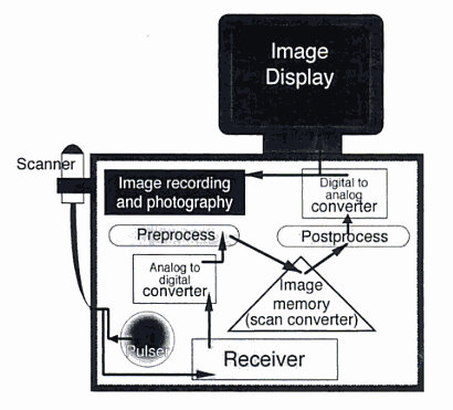
Before we explore what ultrasonography is, let us first examine the terms ultrasonography and ultrasound. According to the AMA Manual of Style, ultrasonography refers to the imaging procedure itself; whereas, ultrasound refers to the sound waves that penetrate the body during ultrasonography. These terms are not synonymous.Animals were first studied using ultrasonography as early as 1949. By the mid-1970s, veterinarians at academic centers were using ultrasonography on clinical patients. Today, the veterinary practice of ultrasonography is nearly ubiquitous. The basic principle of ultrasonography involves the use of high-frequency sound waves (ultrasound waves) to examine the interface between 2 different tissue types—tissues of different densities.
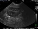
Ultrasound waves are sound waves that exist above the range of sound audible to human beings. They’re produced by electrical stimulation of piezoelectric crystals which are housed in a transducer and aimed at a specific area of the body. These waves travel through the body and are reflected back to the transducer at any point where there’s a change in density. The transducer is connected to an ultrasound machine that determines the intensity level of these “echoes” and maps their location as a static form on a screen (still image). Fluid is mapped as black and various types of tissue are mapped as shades of gray. Alternatively, a transducer can detect shifts in frequency that are proportional to the velocity of a moving object and thus provide a moving (Doppler) image on the screen.
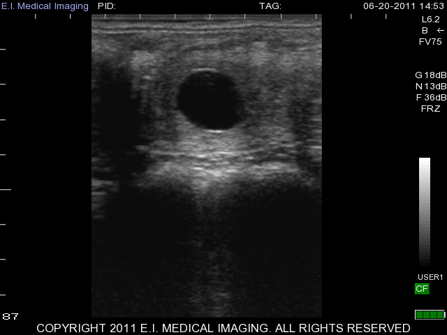
(image: Small Animal Ultrasound by Ronald Green)
Ultrasonography provides the veterinary clinician numerous diagnostic advantages:
• Non-invasive
• Easy and quick to perform
• Portable
• Minimal animal patient preparation (no tranquilizers or anesthesia)
• No ionizing radiation
• No known risks
• Normally no need for animal sedation
• Inexpensive (when compared with computerized axial tomography scan and magnetic resonance imaging)
Disadvantages of ultrasonography include that it’s impossible to image air-containing structures and bone. Furthermore, the quality of the resulting image is dependent on operator skill.
Let’s now examine some clinical uses of ultrasonography in animals.
30 Day Bovine Sheep Pregnancy 15d Equine Canine Liver
Ultrasonography is primarily used to examine fetuses in utero. Furthermore, unlike radiography, ultrasonography can also be used to examine the pancreas, adrenal glands, ovaries, lymph nodes and internal structures of the eye. Many special radiographic procedures including cardioangiography, excretory urography, cystography and upper gastrointestinal contrast studies are now done using ultrasonography. Real-time, Doppler images can be used to examine the bowel and heart. Ultrasonography can also be used to guide procedures such as needle biopsies. Finally, abdominal ultrasonography oftentimes precludes the need for surgery.
So that’s veterinary ultrasonography in a nutshell. .If you have anything to add, please be sure to respond to this post!