Image #1...FEMALE

Image #8, also scanned with an EVO and L7HD probe.What do you see?
Day 4 Image 8
Check back tomorrow morning for the last scan...answers right here tomorrow am!
Here's image #7, scanned with an EVO and L7HD probe. Male or female?
Day 4 Image 7
Check back later today for another scan...answers right here tomorrow am.
Here's image #6, scanned with an EVO and L7HD probe. Can you tell?
Day 3 Image 6
Check back tomorrow am for some more fun!
Have a look at this...#5, scanned with EVO and L7HD probe.
Day 3 Image 5
And #4 is...scanned with EVO and L7HD probe.
Day 2 Image 4
Here's #3—have a look! Scanned with EVO and L7HD probe.
Day 2 Image 3
Don't forget to check back later today for the next one and each day after for new scans. Answers to be revealed Friday afternoon, April 24th!
We will also be posting the images on Instagram @eimedical—follow us there.
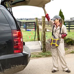
Many equine practitioners who have not come from a sport horse background can be intimidated by the thought of imaging the superficial and deep digital flexor tendons and the suspensory ligament. Getting comfortable with ultrasounding these structures can help you to pay off your equipment faster and provide an important diagnostic option for your clients.
Use a transducer designed for tendon imaging. These probes are higher frequency (and therefore offer finer detail) than a linear rectal probe, for example. The footprint, or size of the imaging window of the transducer, is also smaller, so the structure takes up a larger portion of the monitor. In addition, a tendon probe is ergonomically designed to make tendon imaging easier. A standoff is useful when evaluating more superficial structures, but is not necessarily required for obtaining a good suspensory image.
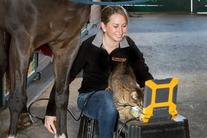
Develop a consistent system. There are several “zone” systems out there; what is important is that you use the same method every time so that you know what your labeling means when archived images are recalled.
Always image distal limbs in two planes, and always image bilaterally. Because tendon areas, for example, will differ among animals of various sizes, the best way to judge pathology in one limb is to compare it with the contralateral one. Save images in longitudinal and cross sections, and label them accurately with zone, measurements, and date.
 For the last nine years, I’ve been proud to work for E.I. Medical Imaging. I’m proud of the tiny but innovative staff that we employ. I’m proud of our dedication to our customers and our investment in developing new and cutting-edge technologies from our small facility in Colorado. I’m proud that we’ve kept the vast majority of our design and manufacturing in the U.S. when it would have been so much easier to outsource everything. I’m proud of the name that we’ve made for ourselves by responding to our customers’ needs over our 30-some years in the veterinary ultrasound industry.
But today, while we as a global community face a new challenge in the wake of the COVID-19 pandemic, I wanted to take a minute and let you know how proud we are of you. Our customer base is made up of animal health experts whose expertise serves us all during this crisis, and many of you are working tirelessly to mitigate the effects of the virus while much of the world retreats into a necessary hibernation.
E.I. Medical Imaging's C9OPU-HD transducer is not your average TVA setup—we were the first to introduce a one-piece probe that’s lighter, slimmer, and easier to clean than the old screw-on clamshell handles.
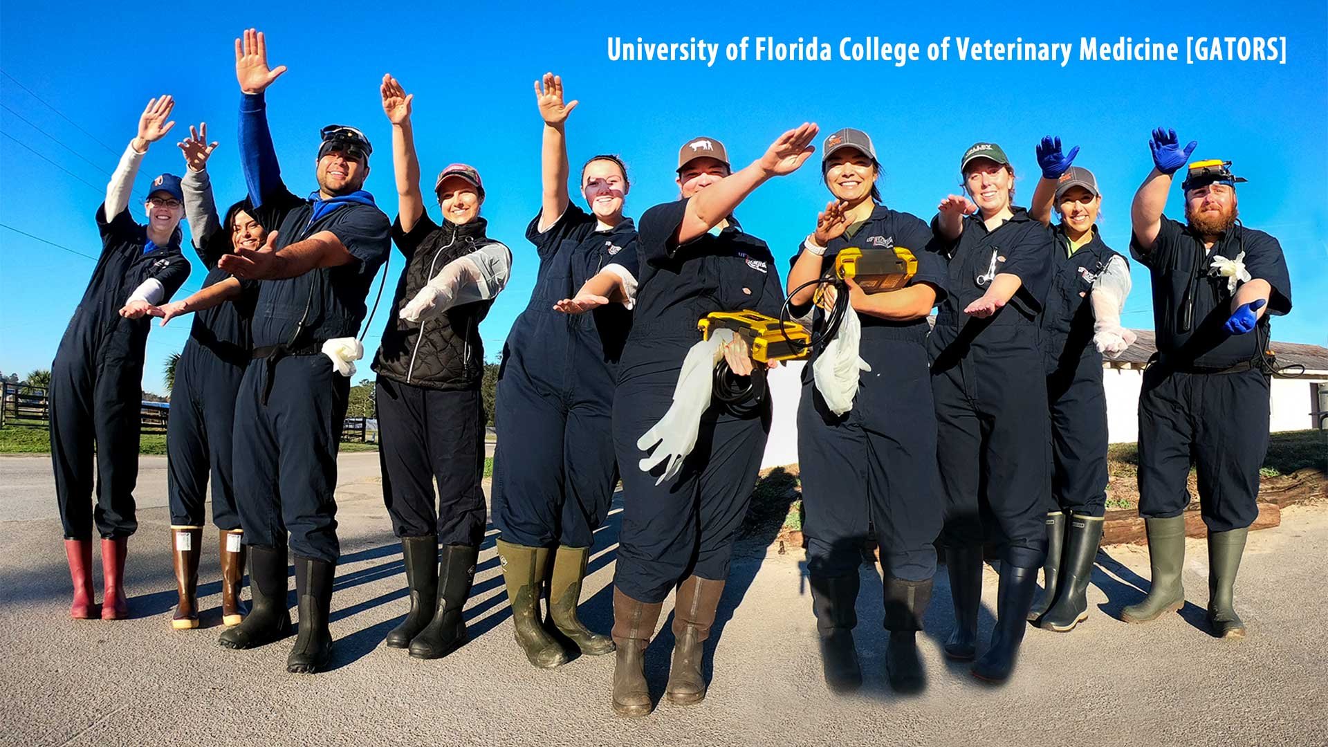
At E.I Medical Imaging we are dedicated to the teaching and development of students. We understand that while in school it may be hard to get hands on experience with ultrasound. That’s why we have launched a program to help prepare you for your future as a veterinarian.
With recently confirmed pregnancies in your broodmare herd, we thought we’d bring you some ultrasound images of equine pregnancies at various stages of gestation...
26-day [L6E transducer]

On a recent bikepacking trip in the UK, E.I. Medical Imaging staff vet Dr. Erika Wierman had this charming encounter...
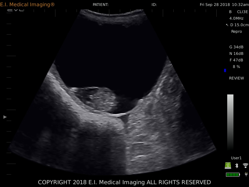
As a veterinarian, you’re trained to understand what you see on an ultrasound machine, but your equine client may be baffled by the images. They may not even know what ultrasound imaging actually is or does, or its value as a diagnostic tool. Here are some tips for explaining ultrasound images to horse owners.
Image: IBEX® EVO® + L7HD, in a Holstein cow
Mummification in bovine fetuses has an incidence of less than 2%. It occurs when there is fetal death for any number of reasons (Trichomoniasis and BVD infections have been specifically implicated as causes), but the CL is retained, the cervix stays closed, and there is no bacteria or oxygen present in the uterus to cause maceration.