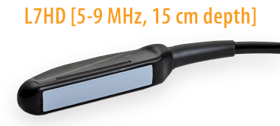
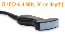
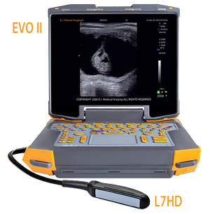
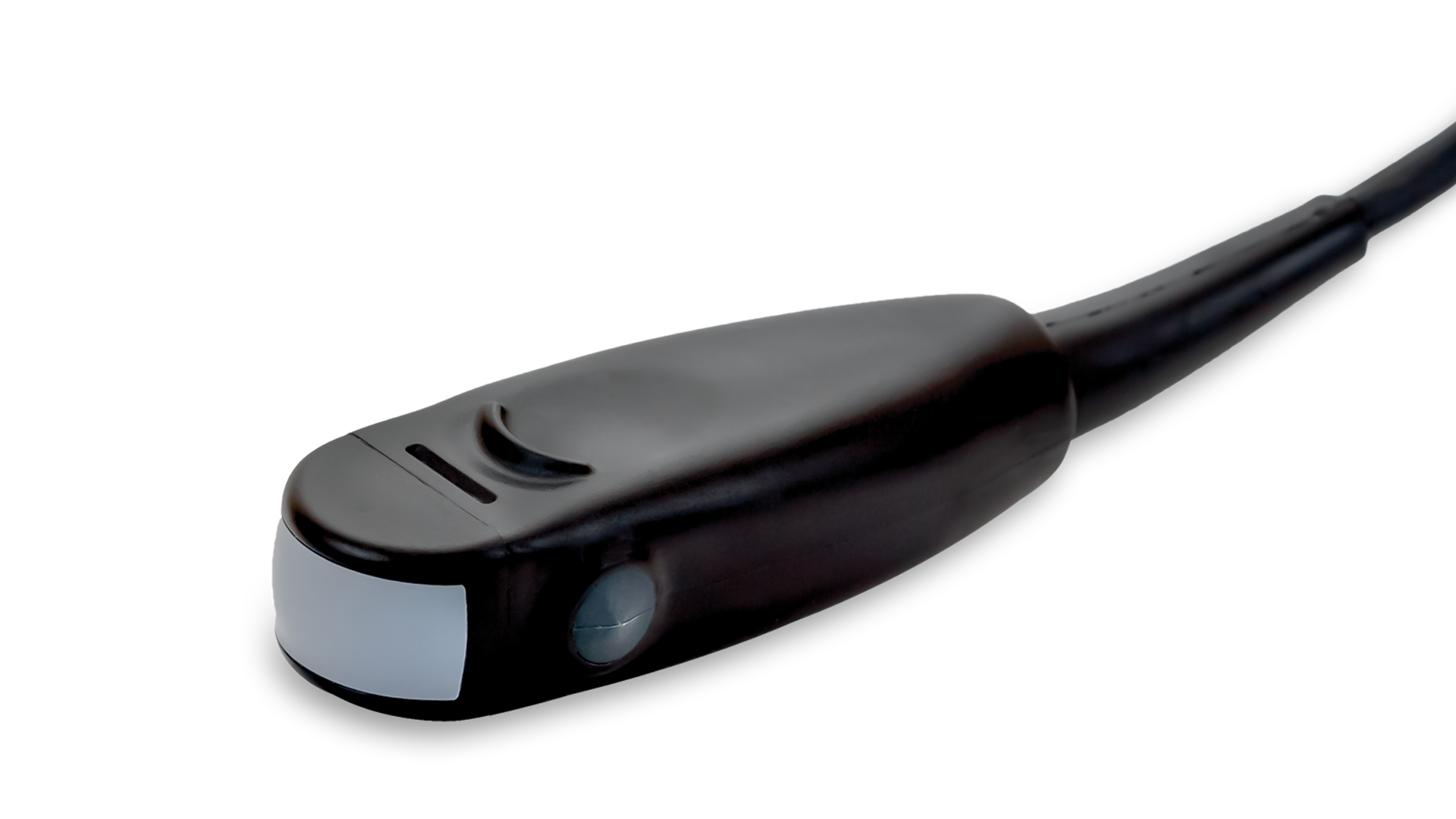
 You’ve probably noticed that the transducers, or probes, on your ultrasound system are named or marked with a number followed by ”MHz”, most likely in the 1-20 range. Often this is how a company advertises their products – for example, a 7MHz linear rectal transducer. Perhaps you’ve wondered what this number refers to or the significance of having a higher or lower number on your probe.
This number (or range of numbers) refers to the frequency of the sound waves produced by that particular transducer. As sound waves travel through tissue, some of them are absorbed or attenuated, and some are reflected back to the transducer to produce an image. Sound waves of a higher frequency are more affected by attenuation, but due to their shorter wavelength are also more accurate in discriminating between two adjacent structures. In contrast, lower frequency sound waves are not as easily absorbed but, due to the longer wavelength, may not discern smaller structures as well.
 What does that mean for you? Transducers with higher frequencies produce a higher resolution image but do not penetrate as well. They are used for imaging small, superficial structures at shallow depths and high resolution. Powerful low-frequency probes are required for imaging at greater depths, although the resulting image may not have the fine detail of one produced at higher frequencies. When operating an ultrasound system, then, it is prudent to select a transducer with the appropriate frequency for the chosen application.
Fortunately, most modern transducers are broadband, which means that they can operate at a range of frequencies. A good rule of thumb is to scan at the highest frequency possible for the penetration that you need to achieve, so that you can optimize the resolution of your image regardless of the depth. Increasing the frequency is a good way to improve the resolution of your image, and decreasing the frequency will help you if you’re struggling to reach deeper structures.
In veterinary medicine, for example, high frequency transducers in the 12-20MHz (~2-6cm depth) range are used to image superficial nerves (regional anesthesia), tendons, and eyes, among other things. Companion animal abdominal and cardiac exams as well as large animal transrectal reproductive exams are typically performed in the 5-12MHz (~6-15cm depth) range, and transabdominal and thoracic imaging of horses, cattle, small ruminants, swine, and even large dogs may be done in the 1-5MHz (~15-30cm depth) range.
For more information on rugged, portable veterinary ultrasound, contact us.


