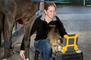
It's Fetal Gender Week!

By Erika Wierman, DVM on Mon, Apr 20, 2020 @ 08:30 AM

Day Four

Image #8, also scanned with an EVO and L7HD probe.What do you see?

Day 4 Image 8              

Check back tomorrow morning for the last scan...answers right here tomorrow am!

Earlier Today...

Here's image #7, scanned with an EVO and L7HD probe. Male or female?

Day 4 Image 7              

Check back later today for another scan...answers right here tomorrow am.

Day Three

Here's image #6, scanned with an EVO and L7HD probe. Can you tell?

Day 3 Image 6              

Check back tomorrow am for some more fun!

Earlier Today...

Have a look at this...#5, scanned with EVO and L7HD probe.

Day 3 Image 5              

Day Two

And #4 is...scanned with EVO and L7HD probe.

Day 2 Image 4              

Here's #3—have a look! Scanned with EVO and L7HD probe.

Day 2 Image 3              

Don't forget to check back later today for the next one and each day after for new scans. Answers to be revealed Friday afternoon, April 24th!

We will also be posting the images on Instagram @eimedical—follow us there.

Continue Reading...

Paying For Ultrasound: Are You Scanning Distal Limbs?

By Erika Wierman, DVM on Fri, Apr 17, 2020 @ 08:30 AM

Portable EVO ultrasound goes wherever you do

Many equine practitioners who have not come from a sport horse background can be intimidated by the thought of imaging the superficial and deep digital flexor tendons and the suspensory ligament. Getting comfortable with ultrasounding these structures can help you to pay off your equipment faster and provide an important diagnostic option for your clients. 

Tips for good, consistent results!

Use a transducer designed for tendon imaging. These probes are higher frequency (and therefore offer finer detail) than a linear rectal probe, for example. The footprint, or size of the imaging window of the transducer, is also smaller, so the structure takes up a larger portion of the monitor. In addition, a tendon probe is ergonomically designed to make tendon imaging easier. A standoff is useful when evaluating more superficial structures, but is not necessarily required for obtaining a good suspensory image.

Scanning with EVO veterinary ultrasound

Develop a consistent system. There are several “zone” systems out there; what is important is that you use the same method every time so that you know what your labeling means when archived images are recalled.

Always image distal limbs in two planes, and always image bilaterally. Because tendon areas, for example, will differ among animals of various sizes, the best way to judge pathology in one limb is to compare it with the contralateral one. Save images in longitudinal and cross sections, and label them accurately with zone, measurements, and date.

Continue Reading...

Equine Follicle Aspiration

By Erika Wierman, DVM on Mon, Feb 17, 2020 @ 08:43 AM

E.I. Medical Imaging's C9OPU-HD transducer is not your average TVA setup—we were the first to introduce a one-piece probe that’s lighter, slimmer, and easier to clean than the old screw-on clamshell handles.

Continue Reading...

Just how portable is your ultrasound equipment?

By Erika Wierman, DVM on Thu, Mar 08, 2012 @ 11:14 AM

PioneerVet.EquineRepro.6.2011.271.jpg

With equine breeding season officially underway, you may be finding yourself re-evaluating your equipment.  Technology changes at lightning speed, and it’s certainly possible that the reliable old ultrasound you’ve been using for years is just pretty darn obsolete now.  While many factors must be considered in the search for a new one, I’d like to take a minute to talk about portability.

Continue Reading...

To learn more about our veterinary ultrasound platforms or to schedule a free demo, click here or call 1.866.365.6596

Trade-in to Trade-up get credit for any ultrasound when you trade up to IBEX premium veterinary ultrasound

Retool to Upgrade breathe new life into out-of-warranty IBEX PRO or LITE to upgrade to our latest technology

Lists by Topic

see all

Posts by Topic

see all

Recent Posts