All-New Patent-Pending eCLi6 Hybrid Rectal Transducer—The Game Changer

By Erika Wierman, DVM on Tue, Mar 12, 2024 @ 10:30 AM

We’ve just launched something truly innovative, and I’m so excited to tell you all about it!eCLi6-3

The new, patent-pending eCLi6 transducer for EVO and SA2 is a hybrid probe for transrectal imaging in large animal species (horses and cattle most commonly). To fully appreciate its development, let’s take a quick step back in time.

Continue Reading...

Three Methods to Improve Broodmare Ultrasound

By Erika Wierman, DVM on Sat, Apr 09, 2022 @ 02:55 PM

pregnant-mare-in-barn-blog-EVO3-[1920]

1...Obtain the best image possible
Take the time to completely evacuate the rectum, and use plenty of lube for contact. If your ultrasound system provides variable frequency transducers, remember to operate at the highest frequency that still allows you enough depth to visualize all the pertinent structures; this will optimize the resolution of your image! You’ll acquire a higher quality (and larger!) ovary image scanning at 6cm than you will at 12cm. In addition, become familiar with your equipment; experiment with gain, contrast, and persistence in order to achieve the image that you desire.

Continue Reading...

EVO Shorts

By Erika Wierman, DVM on Thu, Mar 25, 2021 @ 11:20 AM

EVO Shorts

We've just uploaded several short video tutorials on features commonly used on the unique and powerful EVO veterinary ultrasound. You can find the EVO Shorts playlist on our EIMI University YouTube Channel!

Continue Reading...

High-resolution Transrectal Transducers for Equine Repro

By Erika Wierman, DVM on Tue, Feb 02, 2021 @ 09:02 AM

With the breeding season upon us once again, could it be time to reconsider the status quo with regard to transrectal ultrasound transducers?

Continue Reading...

Happy New Year!

By E.I. Medical Imaging on Thu, Dec 31, 2020 @ 03:30 PM

Happy-New-Year

First of all, thanks for a great 2020! In spite of everything, we've had a great year.

Continue Reading...

Using the EVOStream App

By Erika Wierman, DVM on Wed, Nov 18, 2020 @ 09:30 AM

Detailed instructions on these features and more are found in the owner's manual, found at www.eimedical.com/library.

Continue Reading...

Three Things You Didn't Know You Could Do With Your IBEX PRO

By Erika Wierman, DVM on Thu, Nov 05, 2020 @ 09:15 AM

If you’re anything like me, you barely scratch the surface with regard to using all the features on your gadgets. I know that my iPhone, laptop, and GoPro are capable of all kinds of fun things that I don’t take advantage of. Well, it’s very likely that your veterinary ultrasound also has some hidden gem features that you haven’t used!

Continue Reading...

Paying For Ultrasound: Are You Scanning Distal Limbs?

By Erika Wierman, DVM on Fri, Apr 17, 2020 @ 08:30 AM

Portable EVO ultrasound goes wherever you do

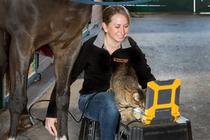
Many equine practitioners who have not come from a sport horse background can be intimidated by the thought of imaging the superficial and deep digital flexor tendons and the suspensory ligament. Getting comfortable with ultrasounding these structures can help you to pay off your equipment faster and provide an important diagnostic option for your clients. 

Tips for good, consistent results!

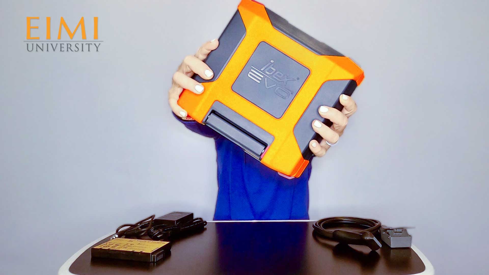
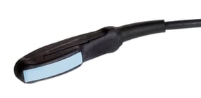
Use a transducer designed for tendon imaging. These probes are higher frequency (and therefore offer finer detail) than a linear rectal probe, for example. The footprint, or size of the imaging window of the transducer, is also smaller, so the structure takes up a larger portion of the monitor. In addition, a tendon probe is ergonomically designed to make tendon imaging easier. A standoff is useful when evaluating more superficial structures, but is not necessarily required for obtaining a good suspensory image.

Scanning with EVO veterinary ultrasound

Develop a consistent system. There are several “zone” systems out there; what is important is that you use the same method every time so that you know what your labeling means when archived images are recalled.

Always image distal limbs in two planes, and always image bilaterally. Because tendon areas, for example, will differ among animals of various sizes, the best way to judge pathology in one limb is to compare it with the contralateral one. Save images in longitudinal and cross sections, and label them accurately with zone, measurements, and date.

Continue Reading...

Equine Fetal Development

By Erika Wierman, DVM on Thu, Aug 15, 2019 @ 08:33 AM

With recently confirmed pregnancies in your broodmare herd, we thought we’d bring you some ultrasound images of equine pregnancies at various stages of gestation...

26-day [L6E transducer]

Continue Reading...

Explaining Ultrasound Images To Your Equine Clients

By Erika Wierman, DVM on Tue, Jul 09, 2019 @ 08:31 AM

Pregnant or not?

2018Sep28-10.32.55
As a veterinarian, you’re trained to understand what you see on an ultrasound machine, but your equine client may be baffled by the images. They may not even know what ultrasound imaging actually is or does, or its value as a diagnostic tool. Here are some tips for explaining ultrasound images to horse owners.

Continue Reading...

To learn more about our veterinary ultrasound platforms or to schedule a free demo, click here or call 1.866.365.6596

Trade-in to Trade-up get credit for any ultrasound when you trade up to IBEX premium veterinary ultrasound

Retool to Upgrade breathe new life into out-of-warranty IBEX PRO or LITE to upgrade to our latest technology

Lists by Topic

see all

Posts by Topic

see all

Recent Posts