Last month I had the pleasure of teaching at the Colorado VMA’s small ruminant meeting, and it got me thinking about some of the common misconceptions and mistakes made when using ultrasound to diagnose pregnancy in sheep and goats. I thought I might take this opportunity to highlight my top five tips.
1 Timing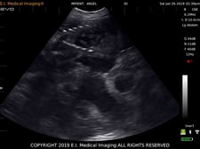 While transrectal ultrasound can be used to diagnose pregnancy as early as 20 days, most exams are done with the non-invasive transabdominal approach. Seasoned practitioners are comfortable with this technique starting around 30-35 days, but many prefer to schedule the initial exam around 45 days. By this time, the fetus and placental structures are more well developed, fetal heartbeats are easy to discern, and the likelihood of missing a pregnancy is significantly lower, but the uterus is still small enough to have a better chance of counting multiples.
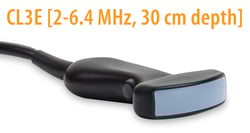
2 Preparation Carefully select the appropriate transducer for the job (see previous blog entry on probe selection here). For example, I personally like the EVO C6E transducer for pygmy and dwarf breeds due to the high resolution and small footprint, but most larger breeds would benefit from a low frequency tool like the CL3E or CLi3E for its power and penetration up to 30cm.
 Make sure you have plenty of contact medium (I usually just use alcohol, but an acoustic gel, OB lube, or even hand sanitizer will work as well). If you are working with a densely-coated breed and want the best images possible, consider having clippers on hand. Sheep often have a thick coating of lanolin in the udder region, so having a towel handy to wipe the waxy debris away will help with adequate contact as well.
3 Positioning
As a rule of thumb, most pregnancy diagnosis is done from a right-sided approach in order to avoid image obstruction by the rumen. Start in the relatively hair-free inguinal area, staying lateral to the udder and scanning medially. The non-gravid uterus or early pregnancy may be found quite far back in the pelvis, sitting just dorsal to the bladder, but later in gestation you will find the fluid-filled uterus moving more cranially. Positioning of the probe can be done from the flank region (a lateral approach) or from between the hind legs, depending upon user preference and facility/restraint design. If a pregnancy is suspected but not visualized, it’s a good idea to scan from the left side as well to make sure a small embryo isn’t missed.
4 Ascertain fetal viability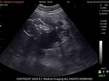 By employing ultrasound early enough in the pregnancy to visualize as much of the fetus or fetuses as possible, you should be able to determine to a reasonable degree the condition of that pregnancy. Look for fetal movement, a consistent fetal heartbeat, and normal, recognizable anatomy. Fluid should be relatively black; the “snowglobe” effect could indicate a higher-than-normal degree of cellular debris within the placenta.
Similarly, ultrasound is incredibly useful for identifying hydrometra or mucometra (“pseudopregnancy,” common in goats)—you’ll see a uterus full of fluid but lacking a fetus and placentomes.
In high risk pregnancies or cases of pregnancy toxemia (ketosis), ultrasound is critical for monitoring for fetal death.
5 Counting fetuses
Many small ruminant owners or producers are very interested in knowing whether their animals are carrying multiple fetuses. This information can be helpful in guiding their nutrition program and preparing for possible dystocias. Despite its many benefits, ultrasound is not the ideal diagnostic tool for this task due to its dynamic nature. Since the transducer is constantly moving, it can be very easy to count the same fetus twice! Unfortunately, radiographs are not practical in most sheep and goats, so we do our best to count with ultrasound. This is best done between 45 and 90 days, but is still not always incredibly accurate. Scan patiently and from both sides, looking for a view of multiple kids or lambs in the same screen. Beware of scanning through a plane that has two parts of the same fetus in it—if you’re seeing a head and hind legs in close proximity, be aware that they may belong to one body. Plenty of practice will make you more confident, but use your counts as a general guideline and don’t rely on them.