A lot has changed since I wrote a blog post for EI Medical in 2011! I have a new last name, two little kids and I no longer use clippers when scanning calf lungs. I am still scanning calf lungs with my Ibex PRO/r and teaching veterinarians and veterinary students the technique. Out on farm and in research, we are now all using one scoring system developed by Dr. Terri Ollivett from University of Wisconsin.
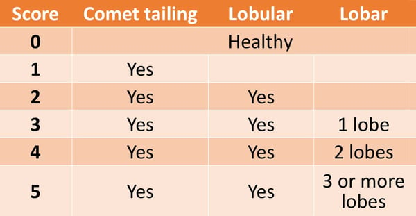
This semi-quantitative scoring system assigns a score depending on the type of lesion(s) observed and extent of consolidation. To count the number of lobes affected for this scoring system, you just need to be confident in your rib space location and corresponding bovine anatomy while scanning. Having one scoring system has been very beneficial as veterinarians and researchers collaborate.
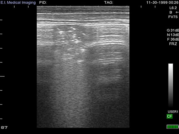
Example of lobular pneumonia and Lung Score of 2, below. Area of consolidation >1cm and does not extend to include a whole lobe [below].
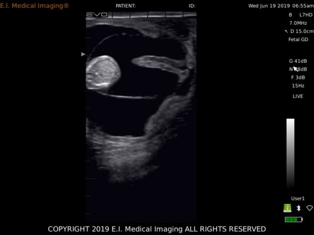
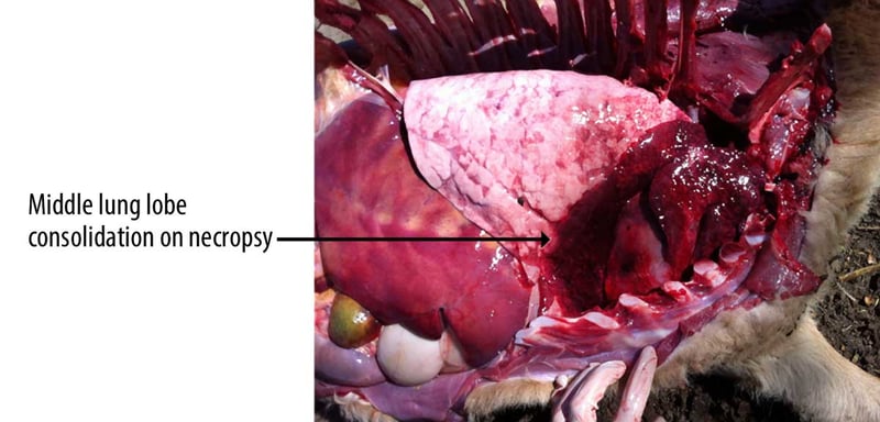
Example of lobar pneumonia, below. Right middle lung lobe (fifth intercostal space) fully consolidated. This is a very common location to see consolidation in calves with bronchopneumonia.
Example of lobar pneumonia, below. Right middle lung lobe (fifth intercostal space) fully consolidated. This is a very common location to see consolidation in calves with bronchopneumonia.Example of lobar pneumonia, below. Right middle lung lobe (fifth intercostal space) fully consolidated. This is a very common location to see consolidation in calves with bronchopneumonia.
As calf raisers focus on quality of the calves they raise, I continue to see more interest in calf lung ultrasound as an objective measure of bovine respiratory disease. Early adopters have been scanning lungs regularly for years and their producers are paying for these services because of the valuable information gathered on the health and future of their calves.
Here are the most common ways I see veterinarians applying thoracic ultrasound in the field:
- Confirming dairy or calf ranch employee’s detection of respiratory disease
- Scanning calves on day 1 of pneumonia diagnosis by on-farm employee
- Scanning calves around day 10-14 of life to confirm if respiratory disease is overlapping with scours
- Animals with unknown respiratory disease history
- Scanning calves at weaning that do not have recorded treatment history
- Scanning calves returning home from a custom calf raiser
- Helping an owner decide which animals should stay in the herd
- Scanning calves with history of 3 or more pneumonia treatments
- Scanning calves at brucella vaccination to determine if animals should stay in the herd
- Scanning calves that are not growing at the same rate as peers
Veterinary students at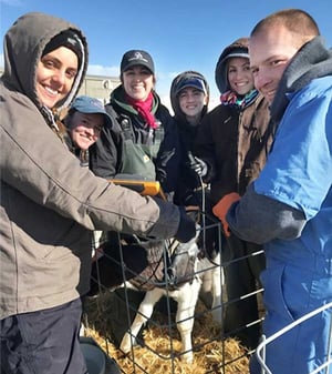
Washington State University
learning to scan lungs
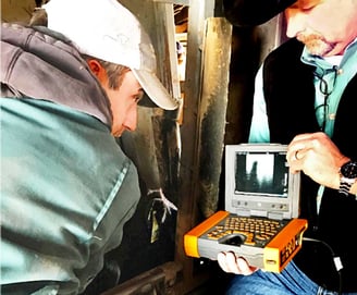 Don’t forget about those native cattle.
Veterinarian scanning beef cattle through a chute.
Dr. Terri Ollivett has written guidelines for dairy calves on timing of when to scan calves in the preweaning period titled #WeanClean. The goal of scanning calves at her different recommended timepoints is to ensure calves are weaned with healthy lungs to greatly reduce the risk of calf pneumonia around this stressful transition time. For more details of this program, visit Dr. Ollivett’s website at
https://thedairylandinitiative.vetmed.wisc.edu/home/calf-health-module/
Calf lung ultrasound is a non-invasive, quick, objective technique to determine the respiratory status of a calf using Dr. Ollivett’s scoring system. Once trained, a veterinarian can scan a calf in less than a minute! Just need the same ultrasound and probe used to preg check cows and 70% alcohol. Remember that calves are prey animals and are very good at hiding their clinical signs of sickness, this is part of why pneumonia continues to be a challenge for the dairy industry. With lung ultrasound, clinical pneumonia can be confirmed, subclinical pneumonia can be detected, and the herd veterinarian can use this valuable information to help producers make more educated decisions on farm.
 Elizabeth Cox MS, DVM
Animal Care Program Manager
California Department of Food and Agriculture
elizms@gmail.com