


Shoot a video testimonial on your phone, GoPro or whatever, telling us how you use your IBEX equipment! Upload it here [up to 100MB]—we'll send a hat and t-shirt to the first 20.
Recent Posts
Measurement and Fetal Aging on the EVO
In our last blog, we looked at fetal aging via ultrasound as a big benefit of imaging over traditional palpation in bovine reproduction.
We've also put together videos on Basic Measurements on the EVO and Fetal Aging on the EVO. Watch how ultrasound can benefit your workflow every day!
Fetal Aging
Fetal aging via ultrasound exam is another big benefit of imaging over traditional palpation in bovine reproduction, as being able to visually assess the pregnancy improves accuracy significantly. Aging is employed in many situations; it can be done to delineate AI from bull-bred pregnancies, to separate animals into calving groups and monitor for dystocias, and to maximize nutritional efficiency throughout the stages of pregnancy, to name a few. While aging via ultrasound is traditionally done prior to 120 days of gestation, we are able to obtain measurements later than ever with the advent of deeper-penetrating, wider field-of-view transducers.
Fetal Anomalies Week—The Answers!
Identification of fetal anomalies or accidents of gestation can not be done in a practical manner during gestation without the use of diagnostic ultrasound, and is one of many examples that demonstrate the superiority of reproductive ultrasound over manual palpation and other manners of pregnancy diagnosis in cattle.
This week we have showcased some of the more common disorders seen in the bovine fetus.
Topics:
early bovine ultrasound
Ibex Portable Ultrasound
how early peg check cows
livestock producers
portable rugged veterinary ultrasound
L7HD
EVO II
bovine reproductive ultrasound
cow herd reproduction
high resolution veterinary ultrasound
ibex veterinary ultrasound
E.I. Medical Imaging
fetal anomalies
Fetal Gender Week—The Answers!
Image #1...FEMALE
Topics:
fetal sexing cows
early bovine ultrasound
Bovine fetal aging using ultrasound
Ibex Portable Ultrasound
how early peg check cows
livestock producers
portable rugged veterinary ultrasound
L7HD
EVO II
bovine reproductive ultrasound
cow herd reproduction
high resolution veterinary ultrasound
ibex veterinary ultrasound
E.I. Medical Imaging
It's Fetal Gender Week!
Day Four
Image #8, also scanned with an EVO and L7HD probe.What do you see?
Day 4 Image 8
Check back tomorrow morning for the last scan...answers right here tomorrow am!
Earlier Today...
Here's image #7, scanned with an EVO and L7HD probe. Male or female?
Day 4 Image 7
Check back later today for another scan...answers right here tomorrow am.
Day Three
Here's image #6, scanned with an EVO and L7HD probe. Can you tell?
Day 3 Image 6
Check back tomorrow am for some more fun!
Earlier Today...
Have a look at this...#5, scanned with EVO and L7HD probe.
Day 3 Image 5
Day Two
And #4 is...scanned with EVO and L7HD probe.
Day 2 Image 4
Here's #3—have a look! Scanned with EVO and L7HD probe.
Day 2 Image 3
Don't forget to check back later today for the next one and each day after for new scans. Answers to be revealed Friday afternoon, April 24th!
We will also be posting the images on Instagram @eimedical—follow us there.
Topics:
fetal sexing ultrasound
twin diagnosis ultrasound
E.I. Medical Imaging
veterinary ultrasound
portable equine ultrasound
portable ultrasound
bovine ultrasound training
ultrasound fetal sexing
Fetal Sexing
Bovine Ultrasounds
Ibex Portable Ultrasound System
reproductive ultrasound
veterinary practice
large animal reproduction
ultrasound for veterinary practice
Paying For Ultrasound: Are You Scanning Distal Limbs?
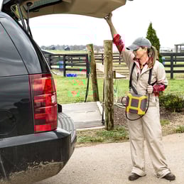
Many equine practitioners who have not come from a sport horse background can be intimidated by the thought of imaging the superficial and deep digital flexor tendons and the suspensory ligament. Getting comfortable with ultrasounding these structures can help you to pay off your equipment faster and provide an important diagnostic option for your clients.
Tips for good, consistent results!
Use a transducer designed for tendon imaging. These probes are higher frequency (and therefore offer finer detail) than a linear rectal probe, for example. The footprint, or size of the imaging window of the transducer, is also smaller, so the structure takes up a larger portion of the monitor. In addition, a tendon probe is ergonomically designed to make tendon imaging easier. A standoff is useful when evaluating more superficial structures, but is not necessarily required for obtaining a good suspensory image.
Develop a consistent system. There are several “zone” systems out there; what is important is that you use the same method every time so that you know what your labeling means when archived images are recalled.
Always image distal limbs in two planes, and always image bilaterally. Because tendon areas, for example, will differ among animals of various sizes, the best way to judge pathology in one limb is to compare it with the contralateral one. Save images in longitudinal and cross sections, and label them accurately with zone, measurements, and date.
Proud of You!
 For the last nine years, I’ve been proud to work for E.I. Medical Imaging. I’m proud of the tiny but innovative staff that we employ. I’m proud of our dedication to our customers and our investment in developing new and cutting-edge technologies from our small facility in Colorado. I’m proud that we’ve kept the vast majority of our design and manufacturing in the U.S. when it would have been so much easier to outsource everything. I’m proud of the name that we’ve made for ourselves by responding to our customers’ needs over our 30-some years in the veterinary ultrasound industry.
But today, while we as a global community face a new challenge in the wake of the COVID-19 pandemic, I wanted to take a minute and let you know how proud we are of you. Our customer base is made up of animal health experts whose expertise serves us all during this crisis, and many of you are working tirelessly to mitigate the effects of the virus while much of the world retreats into a necessary hibernation.
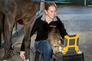
Equine Follicle Aspiration
E.I. Medical Imaging's C9OPU-HD transducer is not your average TVA setup—we were the first to introduce a one-piece probe that’s lighter, slimmer, and easier to clean than the old screw-on clamshell handles.
Student Ultrasound Courses
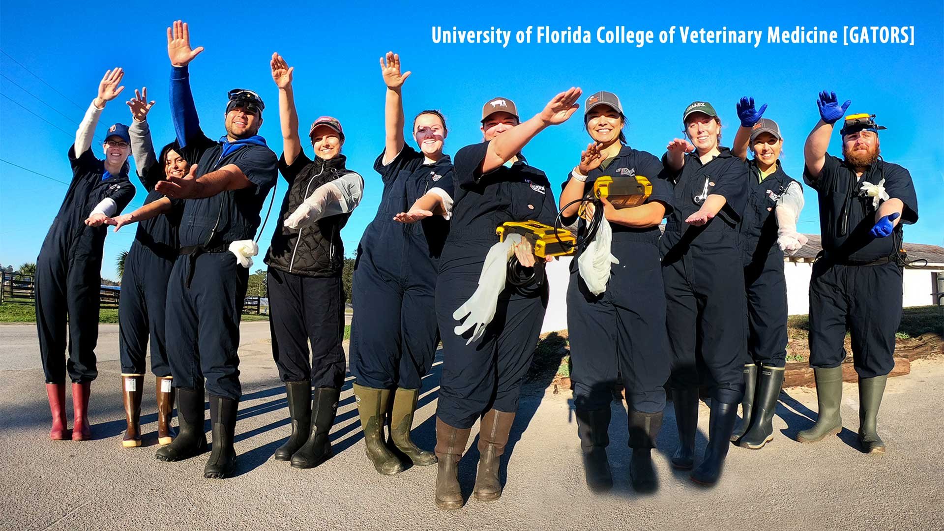
Did you know about our student programs?
At E.I Medical Imaging we are dedicated to the teaching and development of students. We understand that while in school it may be hard to get hands on experience with ultrasound. That’s why we have launched a program to help prepare you for your future as a veterinarian.


