


With recently confirmed pregnancies in your broodmare herd, we thought we’d bring you some ultrasound images of equine pregnancies at various stages of gestation...
26-day [L6E transducer]

With recently confirmed pregnancies in your broodmare herd, we thought we’d bring you some ultrasound images of equine pregnancies at various stages of gestation...
26-day [L6E transducer]

On a recent bikepacking trip in the UK, E.I. Medical Imaging staff vet Dr. Erika Wierman had this charming encounter...
As a veterinarian, you’re trained to understand what you see on an ultrasound machine, but your equine client may be baffled by the images. They may not even know what ultrasound imaging actually is or does, or its value as a diagnostic tool. Here are some tips for explaining ultrasound images to horse owners.
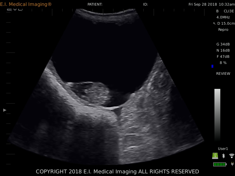
Image: IBEX® EVO® + L7HD, in a Holstein cow
Mummification in bovine fetuses has an incidence of less than 2%. It occurs when there is fetal death for any number of reasons (Trichomoniasis and BVD infections have been specifically implicated as causes), but the CL is retained, the cervix stays closed, and there is no bacteria or oxygen present in the uterus to cause maceration.
Last month I had the pleasure of teaching at the Colorado VMA’s small ruminant meeting, and it got me thinking about some of the common misconceptions and mistakes made when using ultrasound to diagnose pregnancy in sheep and goats. I thought I might take this opportunity to highlight my top five tips.
With our next generation EVO® II and FASTVet™, you can be using ultrasound EVERY DAY in your companion animal practice!
With FASTVet™ techniques, you don’t need to be an expert sonographer to assess and monitor your patients. These are standardized, goal-directed exams that any veterinarian can easily learn and implement. Built-in procedural videos provide immediate assistance in performing FASTVet™ protocols. And in emergencies, no other tool offers such unparalleled, exigent diagnostic assistance.
All ultrasounds have gain control. It’s often a knob, button, and/or a series of sliders on the console, and it’s one of the most used and adjusted scanning parameters... but do you know what it really does?
Most people think of gain as a brightness adjuster, and while it’s true that turning your gain up will brighten the image, it’s helpful to understand how it actually works. Gain is a uniform amplification of the ultrasonic signal that is returning to the transducer after it travels through the tissue. So rather than brightening the monitor, the image on the screen is whitened by a uniform margin, as though the returning signal is stronger than it is, to make it easier to see.
 There are an overwhelming number of transducer (probe) options on the market these days, marketed for different species and applications. What do you need to consider when selecting one? Whether you are shopping for a new system or transducer, or simply deciding which of your current probes to use for a specific purpose, here are a few tips to keep in mind.
 You’ve probably noticed that the transducers, or probes, on your ultrasound system are named or marked with a number followed by ”MHz”, most likely in the 1-20 range. Often this is how a company advertises their products – for example, a 7MHz linear rectal transducer. Perhaps you’ve wondered what this number refers to or the significance of having a higher or lower number on your probe.
If you’re new to ultrasound, an image may look like nothing more than a swirling array of grey tones on the screen. Interpretation requires an understanding of anatomy and physiology, but also of how ultrasound technology functions.
Consider the sonar produced by a bat in flight. The bat emits high frequency sounds, which then bounce off of objects in its proximity and return to the bat. The animal uses the strength, direction, and timing of the returning sound to determine where those objects are and to avoid a collision.


